
What is shockwave therpy?
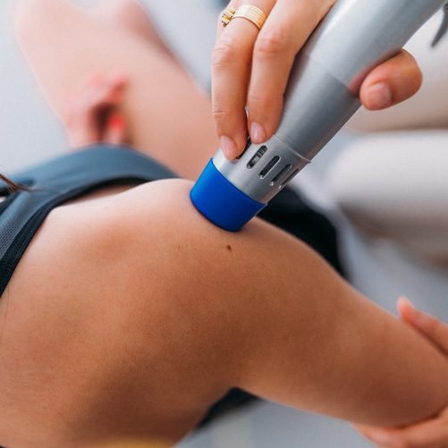
Extracorpuscular Shockwave Therapy (ECSWT) is an FDA cleared medical device that uses high energy acoustic waves via transcutaneous application. This noninvasive therapy uses an electromagnetic motor to create shockwaves. These shockwaves are produced when a projectile is accelerated towards an applicator creating a cavitation or sound wave. Upon impact, a wave is produced which propagates in radial direction from the applicator.
The handheld applicator has the ability to interchange heads, each head has its own property, and can impart varying levels of energy into the body's tissues. Some heads are flat and broad, imparting less energy, and more suited to painful areas, while other heads are convex or pointed and impart higher levels of energy into tissues. There are various diameters of heads as well with the smaller the diameter the higher the energy output, but the least comfortable during therapy, thus a larger diameter head might be needed at the start of therapy to reduce pain during therapy.
HOW DOES IT WORK
The essential principle behind this technique revolves around the action of shockwaves, which are rapid but short duration acoustic waves that carry energy and can propagate through tissues. In general, ESWT bears positive effects in the management of cartilage and bone diseases. This therapy has shown promising results in the treatment of various musculoskeletal disorders, including tendinitis, epicondylitis, plantar fasciitis, trochanteritis, and “jumper’s knee”.It is well known that pressure waves, also called sound waves, are oscillating mechanical waves with the ability to propagate through solids, liquids and gases. As previously introduced, shockwaves are a non-linear type of pressure wave with a short rise time; a shockwave lasts up to 10 μs.The positive and negative phases of shockwaves exert certain effects on interfaces between various tissues and their different densities. In the positive phase, high pressure shockwaves may hit an interface and be reflected or they may pass through and gradually become absorbed. During the negative phase (also referred to as tensile phase), the shockwave generates cavitation at the tissue interfaces, which results in the subsequent formation of air bubbles. The air bubbles then implode with high speed, producing a second wave of shockwaves or micro-jets of fluid.
Cavitation is the formation of vapour bubbles in a liquid wherever the pressure of the liquid falls below its vapour pressure (this is what happens when you crackk your knuckles – the gas in the liquid implodes). This indirect effect occurs in both radial and focused shock waves. These vacuum bubbles induce local shear forces when collapsing at the end of the phase of negative pressure. This cavitation damages the affected tissues.
Capturing images of the cavitation bubbles is not easy but has been performed by Kiessling et al. (2015) and Schlaudraff et al. (2014). Both studies describe the same process which can be seen here.
Although pictures of cavitation are hard to generate radial shockwave machines have been captured creating them. An example is seen below
Radial Shockwave Theapy (RSWT) is described by the diverging pressure field of RSWT devices, which reach maximal pressure at the source instead of selected depths in tissues. Radial shockwaves are not generated in water. Instead, they are generated upon acceleration of a projectile, using and electromagnetic motor which has an extremity connected to an applicator. The projectile is accelerated until it collides with the applicator and, subsequently, the pressure wave that is generated is relayed into the body.
Firstly, in the physical phase, shockwaves cause a positive pressure to generate absorption, reflection, refraction, and transmission of energy to tissues and cells. Additionally, it appears that cavitation increases the permeability of cell membranes and ionization of biological molecules. Secondly, in the physicochemical phase, the physical stimulus leads to biochemical reactions. ESWT triggers the release of biomolecules such as adenosine triphosphate (ATP) for the activation of cell signaling pathways. Thirdly, the chemical phase has shockwaves altering the functions of ion channels in cell membranes and mobilization of calcium. Lastly, the biological phase is where ESWT plays its role in modulating angiogenesis, anti-inflammatory effects, and healing of bone and soft tissue wounds. In addition to the effects previously discussed, ESWT-induced neovascularization and improvement of blood flow may also potentialize the regenerative properties of this technique. It has been hypothesized that the biological effects of ESWT are a consequence of mechanotransduction, a phenomenon which relies on the action of ultrasonic vibrations on tissues, which then lead to regeneration and healing. So far, there are two principal hypotheses proposed to explain the analgesia induced by shockwave (SW) treatment. One of them suggests that SWs degenerate nerve fibers from small immunoreactive neurons, therefore decreasing the concentration of pro-inflammatory mediators. The second mechanism is theorized to cause analgesia via hyperstimulation, indicating that SWs trigger the release of endorphins and other analgesic molecules by activating the descending inhibitory system.
In conclusion this non-invasive, medical procedure is capable of triggering cellular and molecular alterations that assist the regeneration of injured tissues. ESWT is mainly responsible for: pain relief, by acting directly on nerve fibers; tissue regeneration by stimulating vascularization; and reduction of calcium deposits in tissues. Numerous publications have proven the efficacy and safety of both radial and focused ESWT for the treatment of many musculoskeletal disorders, including osteoarthritis and different types of tendinopathies. ESWT should be considered in severe cases where conventional treatments prove to be of little success, especially in patients who prefer non-operative alternatives.
Conditions Treated
Plantar fasciitis.
Achilles tendonopathy.
Retrocalcaneal bursitis.
Lateral epicondylosis (tennis elbow).
Medial epicondylosis (golfer’s elbow).
Calcific tendonitis (supraspinatus tendon, etc.).
Patellar tendinosis (jumper’s knee).
Morton’s neuroma.
Chronic stress/non-union fractures.